Scan you believe it? The value of CT and MRI scans in turtle rehabilitation
Over the years, our Turtle Conservation Centre has rehabilitated hundreds of endangered turtles, each with a unique story. Many of the subadult turtles are rescued in dire condition, suffering from a range of complex illnesses like lung disease, septic arthritis, fractures, and more. These issues often require extensive medical treatment to be resolved, and our Turtle Conservation Centre team is committed to giving their patients the best possible care to return them to full health.
In recent years, our team has had the immense privilege of working with Winelands Radiology, allowing our veterinary team to use their Computed Tomography (CT) and Magnetic Resonance Imaging (MRI) services for many of our turtle patients. This has changed the game for our veterinary team, allowing them to elevate their diagnostic capabilities and consequent turtle care to new heights.
Let’s learn more…
How does the team usually visualise the internal structures of our rescued turtles?
Each turtle admitted to the Turtle Conservation Centre after being rescued undergoes a thorough assessment to evaluate their overall health and identify any potential issues that may affect their rehabilitation. This includes a physical exam, radiographs, and blood tests. The information gained from these various steps allows our veterinary team to get a good idea of the turtle’s general health status.
Radiography (commonly known as X-ray imaging) is one of the primary tools our team uses during this admission process. They provide an accessible and cost-effective method to visualise internal structures. “X-rays are very useful for diagnosing conditions of the bones and joints, like fractures or arthritis. It is also a useful tool to evaluate lung health and look for foreign material in the turtle’s body cavity," says Dr Bernice van Huyssteen, our turtle veterinarian.
However, the amount of information gained from X-rays is limited. An X-ray provides a 2D picture of moderate detail but obscures a lot of internal complexity. This makes it difficult to differentiate between various soft tissues, meaning it is more complex to map and track conditions like lung infection. There is also a delay in the development of bone changes and their being visible on X-ray, which can cause certain conditions to be missed during initial assessment.
In many cases, more detailed imaging is required to fully define a condition and formulate an appropriate treatment plan. This is where Winelands Radiology comes in…
Radiology changes the game!
X-rays are a useful, but somewhat limited, way to view a turtle’s skeletal structure and organs. However, CT and MRI take this to a new level!
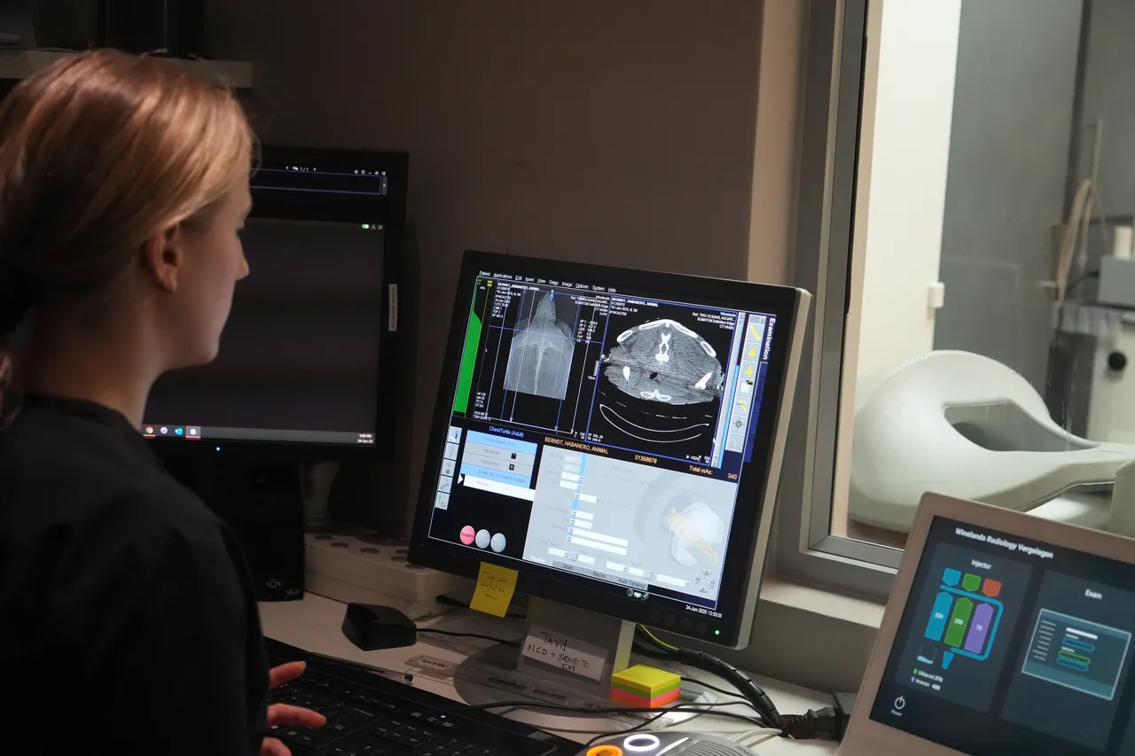
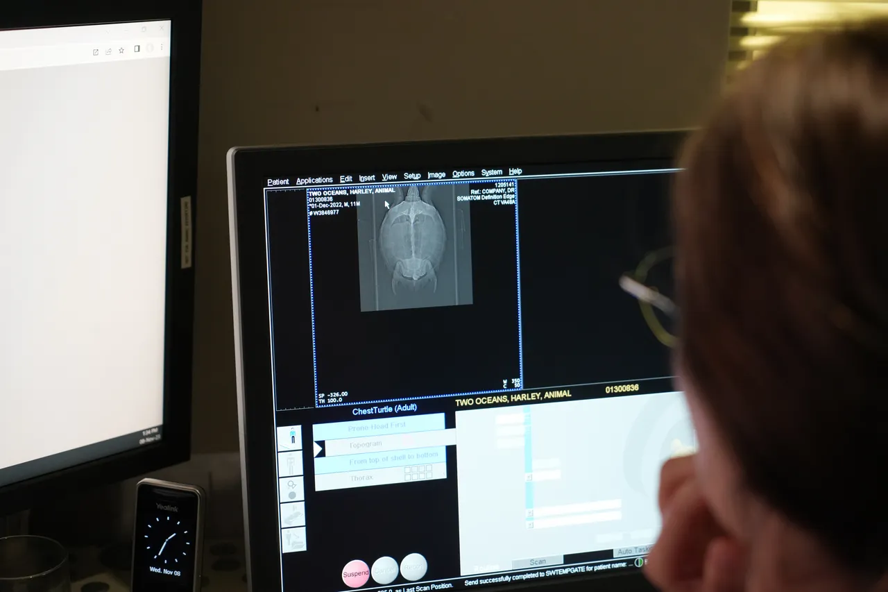
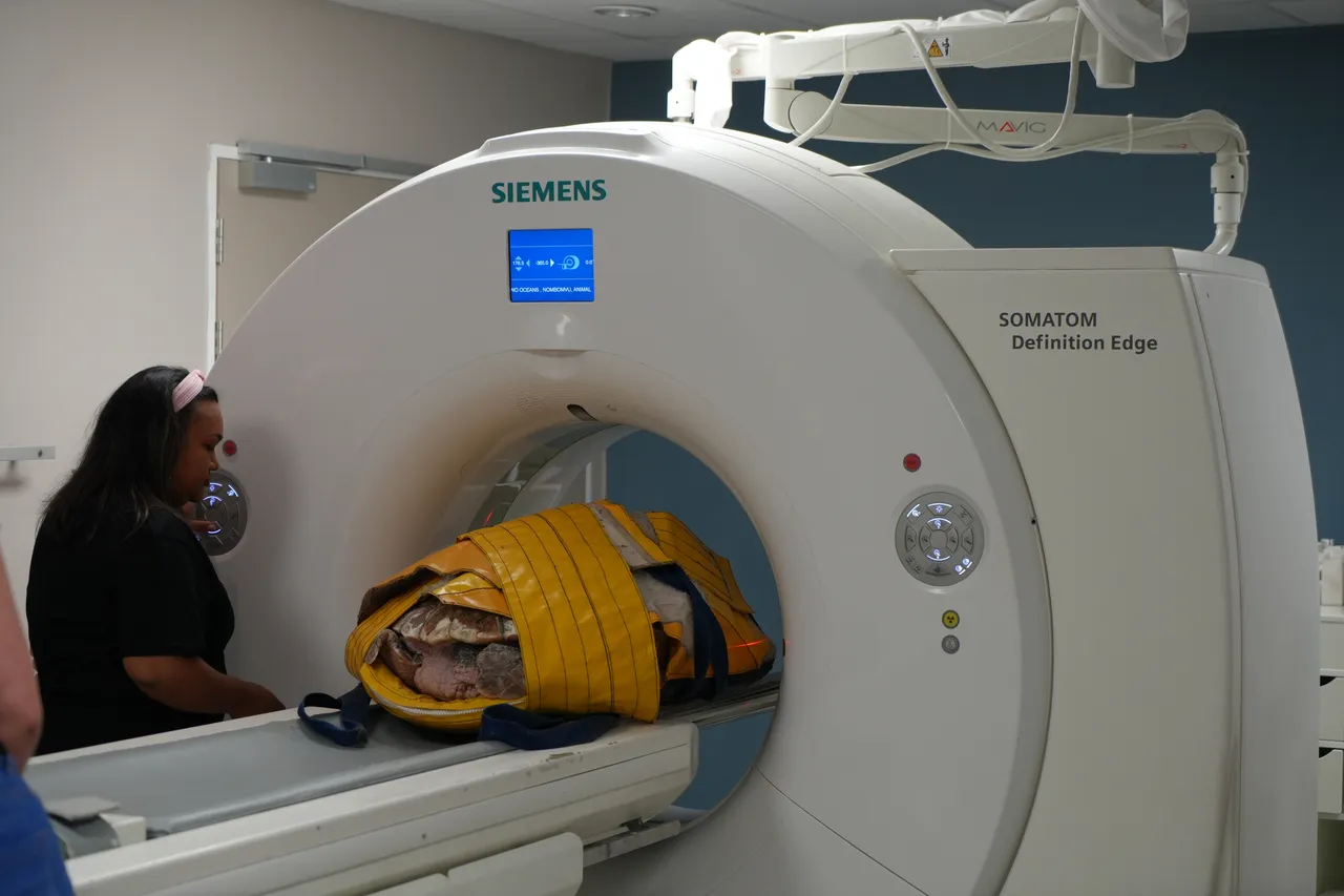
CT scans actually use X-ray technology to create cross-sectional images with incredible detail. These scans are ideal for detecting hard-structure problems, such as minute bone fractures, blood clots, or organ injuries. MRI uses magnetic fields and radio waves (not radiation) to examine soft tissues, like the brain, spinal cord, ligaments, and tendons. These types of scans are particularly useful for animals like turtles, whose rigid shells and overlaying, high-density organs make it difficult to detect subtle internal issues with X-rays.
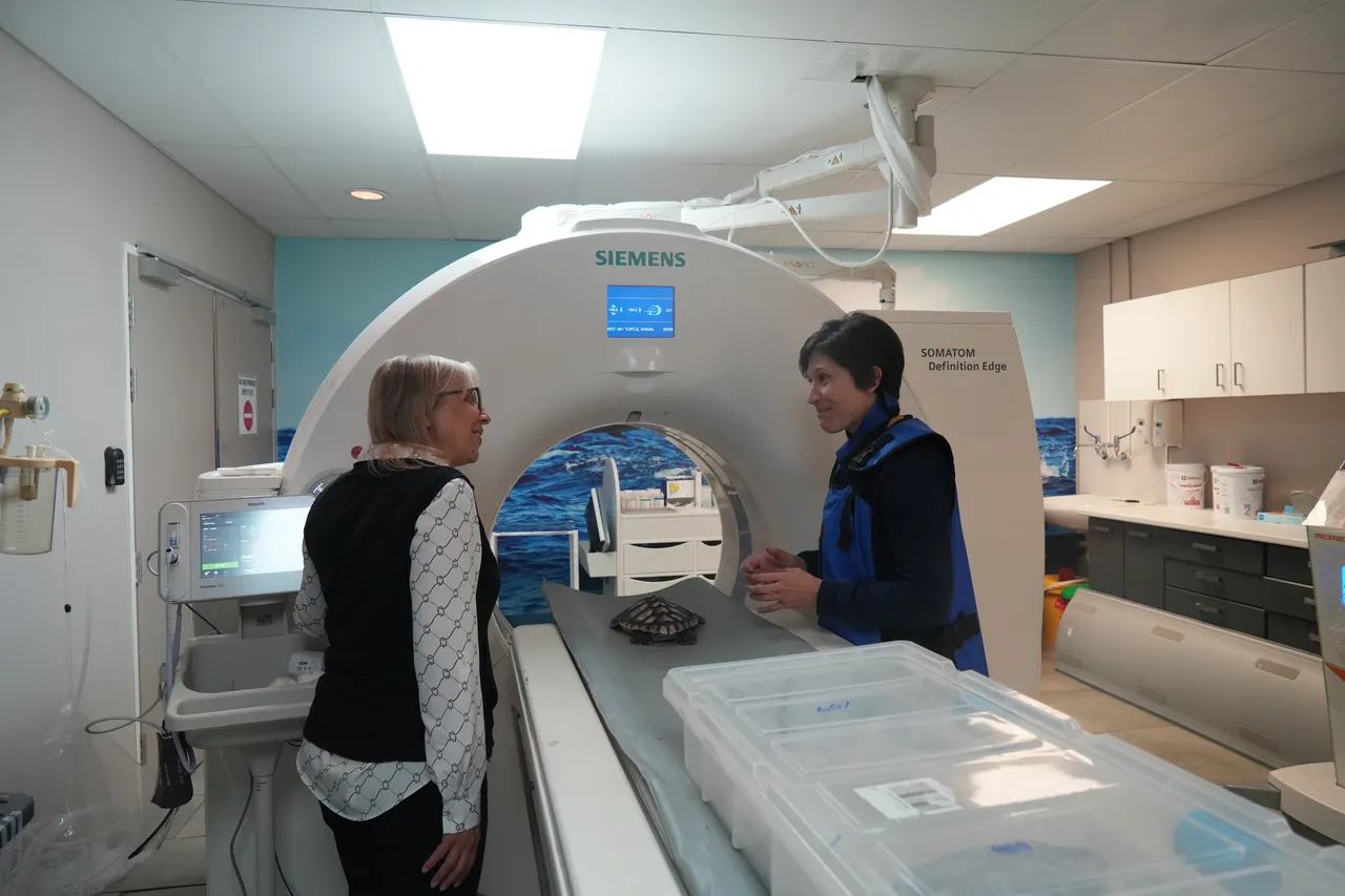
In 2021, radiologist Dr Peter Berndt and his colleagues at Winelands Radiology reached out a helping hand to the Turtle Conservation Centre, offering their equipment and expertise for a few unconventional patients. Peter became known to our team as "turtle dad" for his generous and compassionate work with numerous turtles undergoing rehabilitation at the Turtle Conservation Centre. With Peter and his team's help, we have discovered and successfully treated turtles for issues like respiratory tract infections, lung infections, arthritis, and osteitis.
“Our relationship with Winelands Radiology has completely changed the playing field in the medical care we can offer our turtles. Regular access to CT and MRI has allowed us to diagnose complex conditions such as a pneumocoeloem (an air pocket in the lung that causes buoyancy issues), granulomatous lung disease, tubal bronchiectasis, and map the extent of shell fractures, brain trauma, and related internal injuries, just to name a few,” says Dr Bernice.
Not only do CT and MRI help our vets more accurately diagnose complex conditions, but they are also crucial tools in monitoring each condition’s progression and the turtle’s response to treatment. “These tools have enriched the quality and accuracy of our medical care and directly benefited our turtles’ rehabilitative success,” says Dr Bernice.
How have CT and MRI scans helped our turtles?
Habanero, the loggerhead turtle
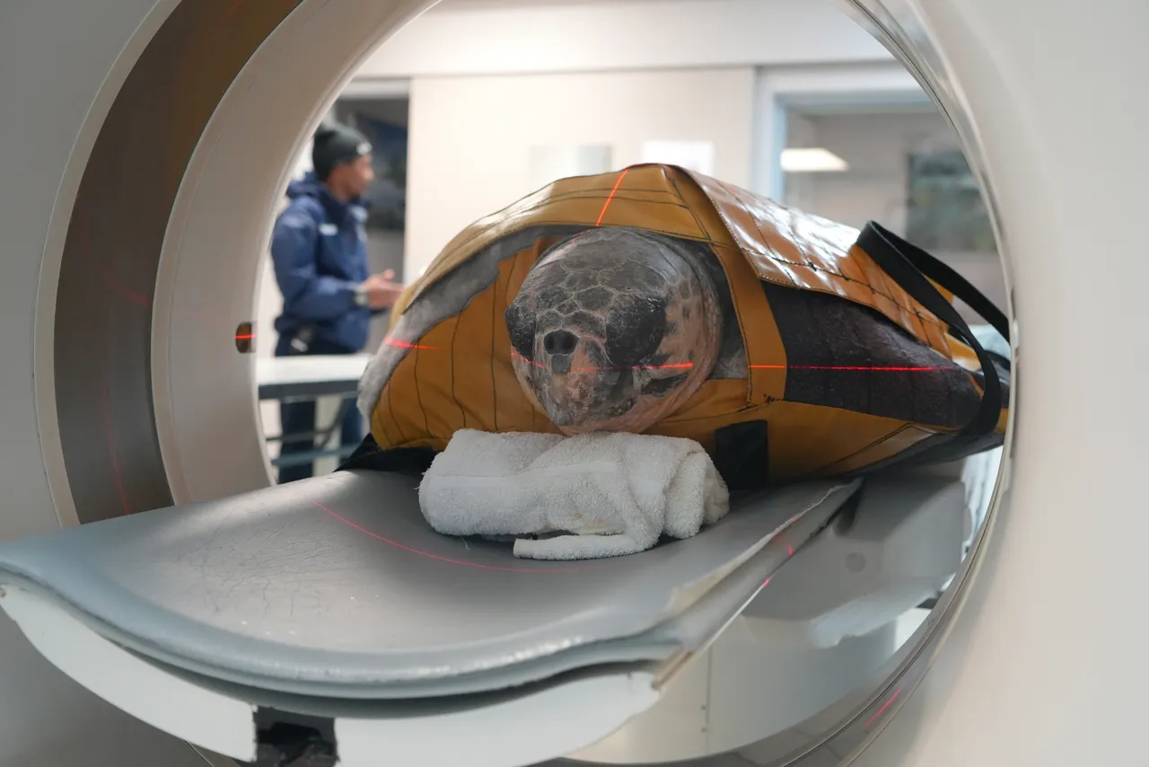
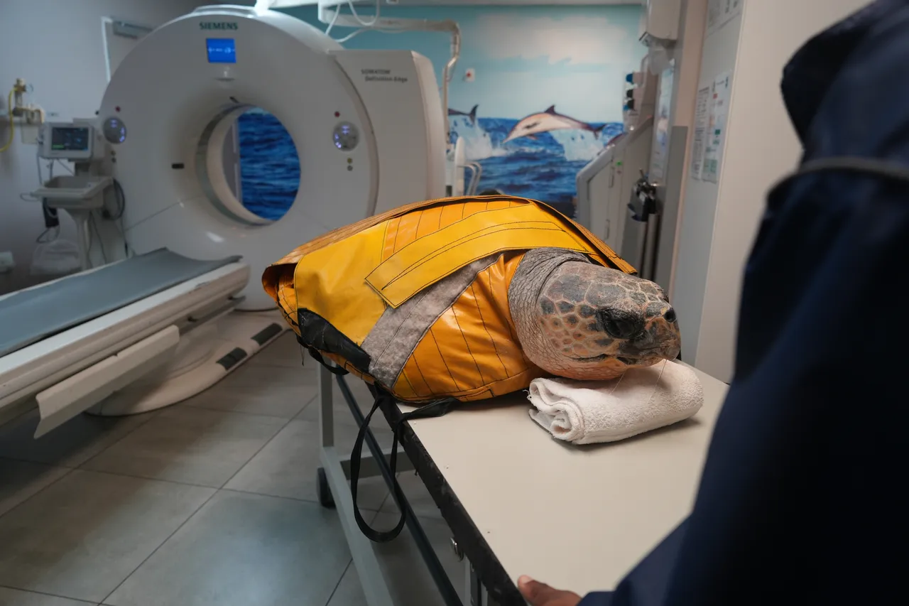
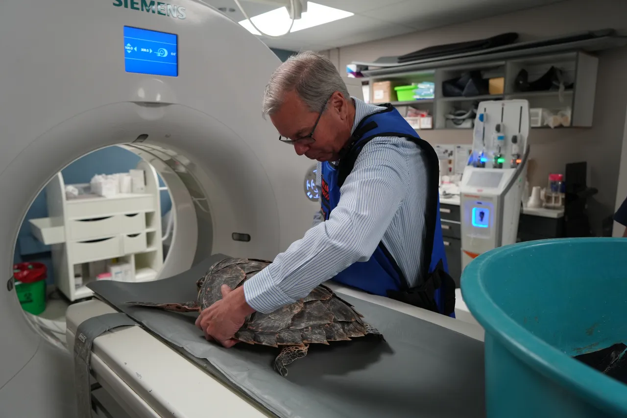
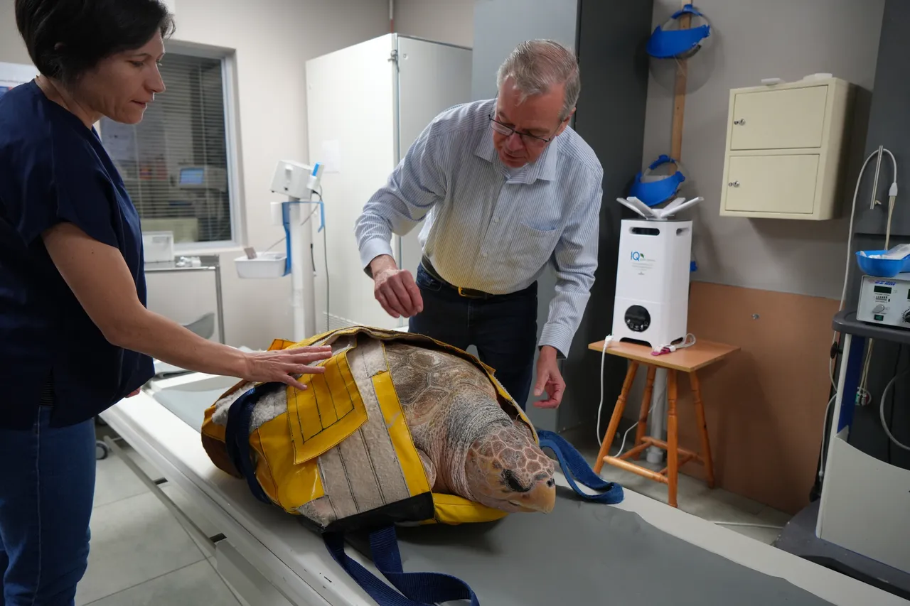
One such patient whose rehabilitation journey has been shaped by access to CT and MRI is Habanero, a large adult loggerhead turtle.
Habanero was admitted to the Turtle Conservation Centre in February in a weak, dehydrated state and covered in marine leeches. A swollen and painful left elbow was obvious on initial examination, and X-rays revealed degradation of the bones in the joint.
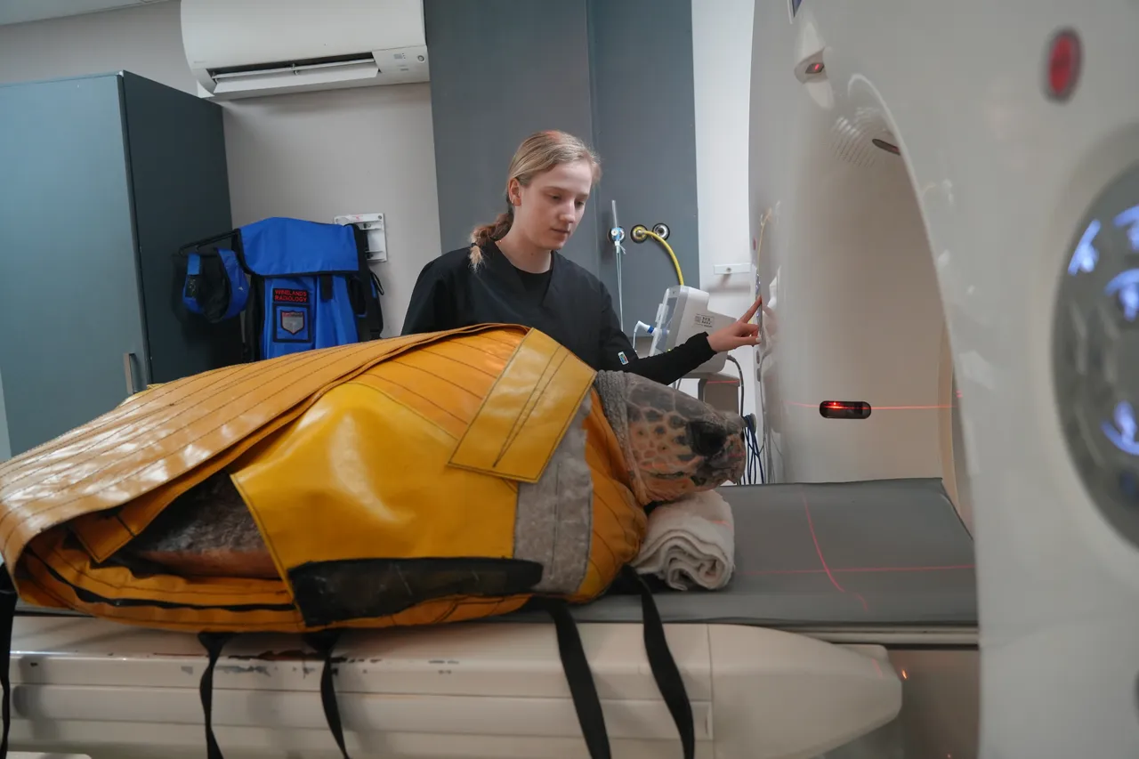
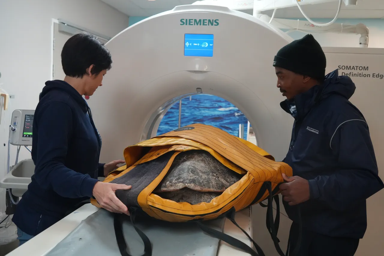
Our veterinary team decided that this issue made Habanero a prime candidate for a CT scan, so off he went to Winelands Radiology, accompanied by our dedicated turtle team and a few other turtle patients. Habanero was a perfect patient, calmly resting in the CT scanner while the Winelands Radiology team expertly worked.
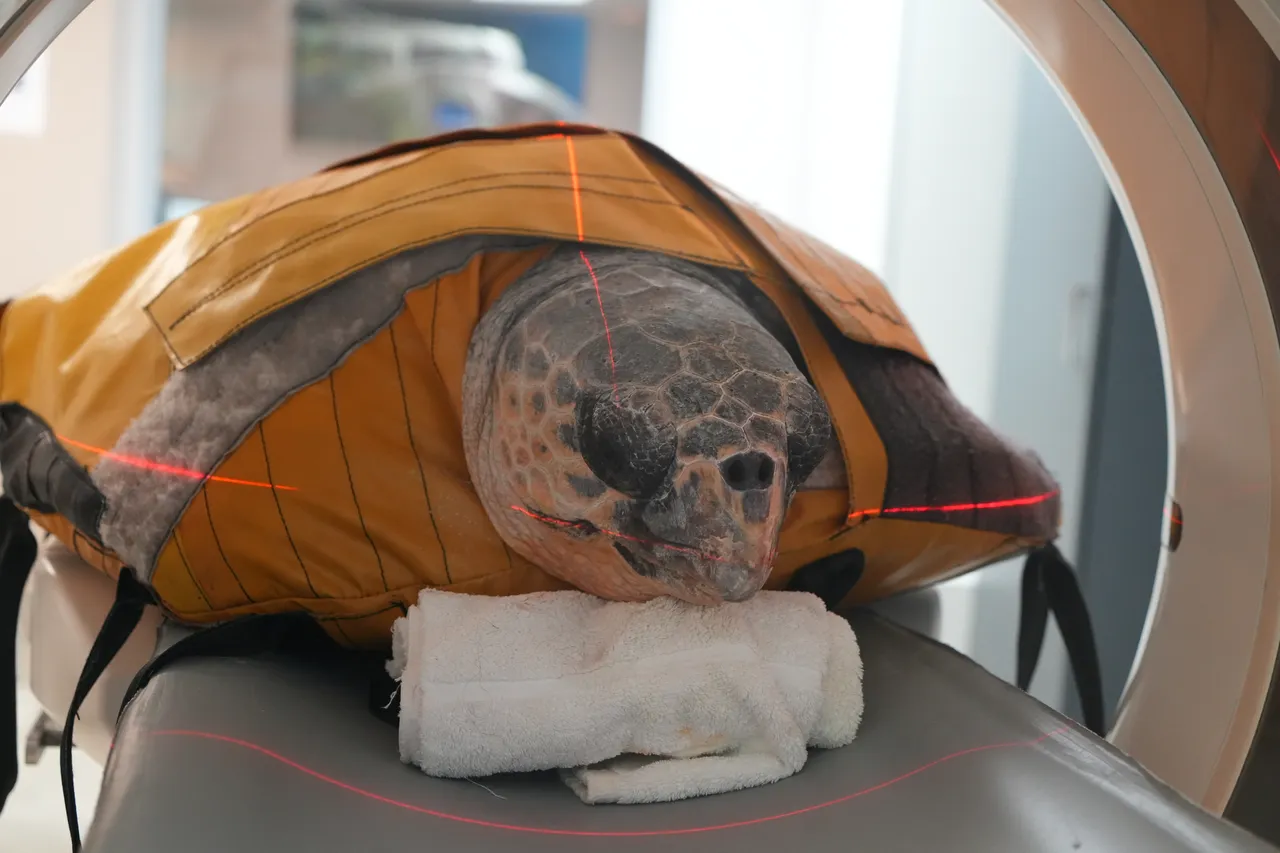
The results from the CT scan confirmed that Habanero had severe septic arthritis in his left elbow. A large amount of necrotic tissue was also identified in the joint, something that could not be identified or quantified based on X-ray imaging alone. Given the extent of damage and necrotic tissue visualised on CT, it was evident that antibiotic treatment alone would not resolve this problem. Surgery would be required to remove the dead and infected tissue before a resolution of the infection could be expected. This highlights the impact and insight offered by CT scans.
Once Habanero was deemed stable enough to undergo an anaesthetic, he underwent surgery to debride the infected joint of all the dead tissue. This procedure, led by Dr Bernice, proved incredibly successful – Habanero recovered well and later returned to Winelands Radiology for a follow-up scan to evaluate the response of the joint post-surgery.
Habanero still has a long rehabilitation journey ahead, but Dr Bernice is hopeful that the joint infection will be successfully resolved. In the meantime, he is progressing well and was even introduced to the I&J Ocean Exhibit to help him gain strength!
Stella and Pebbles, green turtles
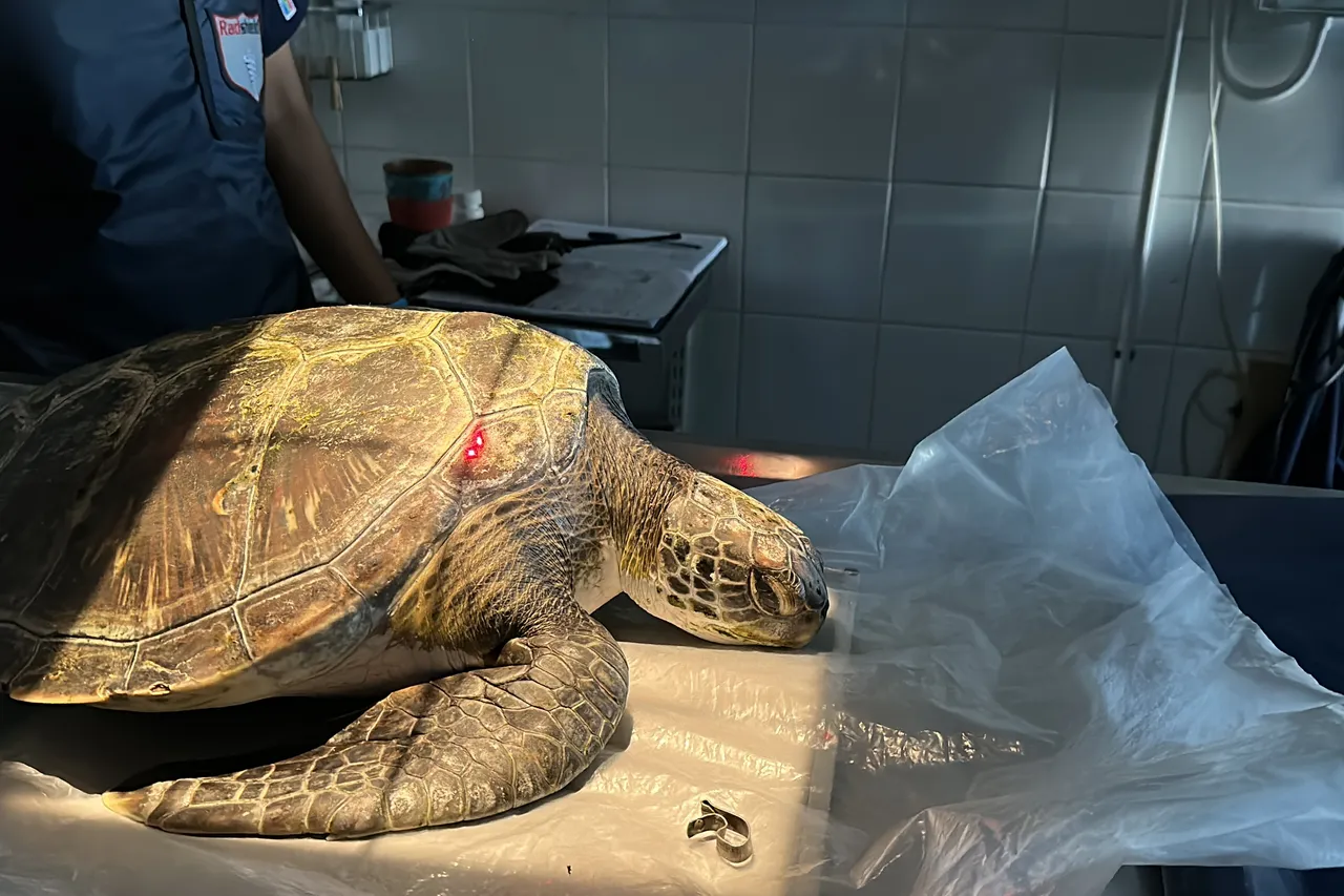
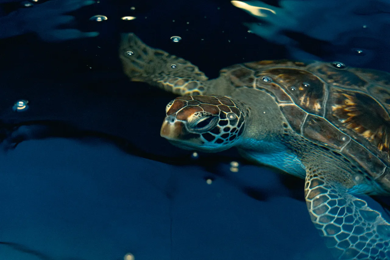
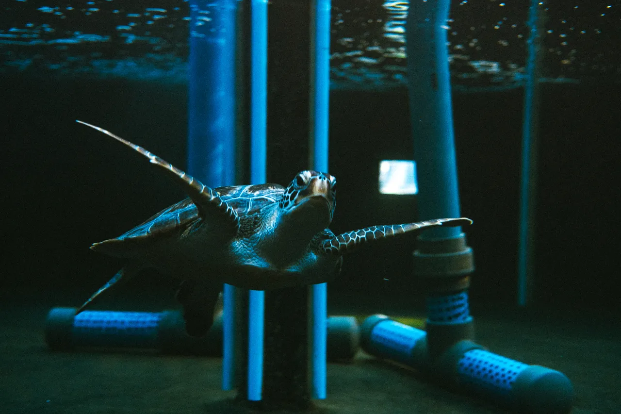
Two other examples of the magic of CT and MRI are juvenile green turtles, Stella and Pebbles.
Both turtles were rescued towards the end of 2024 and have been undergoing treatment for chronic bronchopneumonia (a type of pneumonia in the lungs). Regular CTs at Winelands Radiology allowed our veterinary team to monitor the response to their antibiotic treatment and adjust it accordingly as needed. Finally, after more than a year of treatment, their latest CT scan revealed clear lungs. This meant Stella and Pebbles were given the all-clear to be taken off treatment by Dr Bernice.
Their next CT scan will take place once the turtles have been off antibiotic treatment for two months. “We’re crossing our fingers that the scan will show that they are finally ready for release!” says Dr Bernice.
Turtle rehabilitation can be complicated, with many twists and turns along the way. Each turtle’s story is different, and our turtle team is committed to giving each patient the best treatment they need to return to the ocean. We are privileged that this world-class care includes access to CT and MRI scans with Winelands Radiology. These diagnostic tools, together with the compassionate efforts of the team at Winelands Radiology, have helped get many of our turtles back on their flippers.
Related News

Sign up to our Newsletter
Receive monthly news, online courses and conservation programmes.